
 According to the latest research by Albert Einstein College of Medicine of Yeshiva University, the researchers linked areas of brain injury to particular modified mental processes apparently caused by concussions. Whether concussions occur from an accident, sporting event or a fight, it can supposedly lead to permanent loss of higher level mental processes. Scientists have pondered over for several years whether concussions cause structural damage to brain tissue. Also whether physiological changes that only harm the way brain cells work, explain this loss seems to have been of particular interest.
This research apparently reveals that concussions involve brain damage. The findings propose that diffusion tensor imaging (DTI) could help in detecting concussions and in examining the effectiveness of treatments. DTI is the brain scanning method which is used by the Einstein scientists.
“DTI has been used to look at other brain disorders, but this is the first study to focus on concussions. It proved to be a powerful tool for detecting the subtle brain damage that we found to be associated with concussions,” commented Michael Lipton, M.D., Ph.D., associate director of the Gruss Magnetic Resonance Research Center (MRRC) and associate professor of radiology, of psychiatry and behavioral sciences, and of neuroscience at Einstein and lead author of the research.
Currently, diagnosis for concussions is claimed to be based principally on the accident history of the patient and clinical examinations that check for common symptoms such as headache, dizziness and behavioral abnormalities. Apparently there is no way of knowing, even from these tests, as to who will recuperate rapidly and who will suffer severe consequences. As per the Einstein research, to diagnose concussions, DTI scanning seems to supply an objective method. It can supposedly be used to find out whether brain injury has actually happened following trauma and also to possibly predict the permanent loss of executive function which refers to the capacity to make decisions, organize, set priorities and manage time.
The research included 20 people who have suffered concussions (18 from motor vehicle accident and 2 from falls) and 20 people who were perfectly healthy control subjects. The patients were selected from one hospital emergency room. Apparently 2 of them had lost consciousness for a few minutes. The patients as well as the healthy subjects went through conventional brain imaging with magnetic resonance (MR) and computed tomography (CT) scanning, and a series of neuropsychological tests to examine executive function, which is often damaged after a concussion. Within 2 weeks of their accident, all concussion patients went through brain imaging and testing.
The veteran reviewers who assessed the conventional MR and CT images of patients and controls supposedly discovered no abnormalities in either group. But the neuropsychological results displayed that apparently in executive function, the patients performed more badly than the controls.
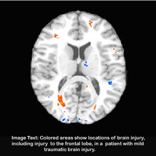
Patients and controls went through diffusion tensor imaging (DTI). It is a recently developed MRI-based imaging technique that can identify understated changes in the brain by calculating the diffusion of water in the brain’s white matter. Abnormal areas were exposed in 15 of the concussion patients but no abnormal regions were spotted in controls. Moreover, the presence of key regions of structural damage in concussion patients could foretell low scores on their executive-function tests. These damaged areas were supposedly placed mostly in the brain’s prefrontal cortex, which is vital for normal executive function and is vulnerable to injury in concussion.
Lipton observed “The problems in functioning caused by concussions often don’t become evident until weeks or months after the injury, suggesting that the brain pathology may actually expand over time. By detecting brain injury early with DTI and then initiating cognitive rehabilitation therapies for those patients, we may be able to limit the effects of concussions.”
Craig Branch, Ph.D., a co-author of the research and director of the Gruss MRRC commented “We are really excited by these findings. For the first time we appear to be able to identify the subtle pathology sometimes caused by concussion, providing researchers a ‘pathology target’ for the development of therapies to reduce or eliminate the damage identified by this novel imaging method.”
Lipton is of the opinion that application of DTI could prompt doctors to start the treatment before time, when it is apparently expected to be most useful. Adding to it, Lipton mentioned that DTI could be useful in estimating the effectiveness of existing therapies for concussion.
This research was published in the August Issue of Radiology.