
 Around last month we reported that men with low-risk of prostate cancer receive aggressive treatment. Now, experts from Rutgers are developing methods that may precisely evaluate the severity of prostate cancer. This is done by examining magnetic resonance images and spectra of a patient’s prostate gland.
This analysis may help physicians identify patients who require aggressive treatment. It may also help to delay or avoid invasive biopsies in patients with low-grade tumors. These findings were achieved by conducting computer analyses of the images and spectra made on 19 patients.
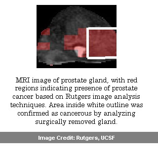
Based on earlier studies at Rutgers, this analysis used powerful, high-resolution magnetic resonance imaging technology. Experts collected prostate gland images from 19 patients who later had radical prostatectomies. They evaluated traditional MR images which offered two-dimensional pictures of the gland’s cellular structure. In addition, they also examined MR spectroscopy which maps concentrations of specific chemicals to areas in the prostate gland. Deviations in concentrations of these chemical metabolites, choline, creatine and citrate highlight the occurrence of cancer.
“The breakthrough we’ve had in the last few months is that we see image signatures that distinguish aggressive cancers from less aggressive ones,†commented Anant Madabhushi, associate professor of biomedical engineering at Rutgers and a member of The Cancer Institute of New Jersey (CINJ).
Scientists compared the MR images and spectra to digital images of the actual excised glands. This was identified as having high-grade or low-grade tumors utilizing the Gleason Grading System. They further used pattern recognition techniques to highlight characteristics of areas in the MR images and spectra that were associated to the cancerous tissue in the excised samples. This was conducted by adopting computerized tools to line up the MR views with digitized images of tissue slices, and to match the diverse resolutions of the images and spectra.
The main purpose is to instruct the computer system to precisely and constantly identify image patterns that relate to several grades of cancerous tissue without having the tissue samples to physically validate. Experts share that these methods have to be conducted on more people before it can be considered for clinical purpose. Presently clinical diagnoses are based on PSA levels in blood, physical examinations and needle biopsies.
Biomedical engineering graduate student Pallavi Tiwari will present these findings at the Medical Image Computing and Computer Assisted Intervention Conference in Beijing, China, on Sept. 22.