
 Detailed analysis may be essential for patients examined by MRI and it claims to be the only way to attain vibrant images for diagnosis. Now, Max Planck researchers from Göttingen have efficiently succeeded in lowering time required for recording images to just one fiftieth of a second.
The mechanism of organs and joints can be recorded, in addition movements of the eye and jaws, bending of the knee and beating of the heart can be filmed. The new, innovative MRI method puts forth essential information about diseases of the joints and the heart. MRI examinations may become more simple and convenient for patients.
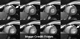
Real-time MRI of the heart is with evaluation time of 33 milliseconds per image and 30 images per second. The spatial resolution is around 1.5 millimetres in the image plane. The eight consecutive images highlight the movement of the heart muscle of a healthy subject for duration of 0.264 seconds during a single heartbeat. The images range from the systolic phase to the diastolic phase. The vibrant signal in the heart chambers is the blood.
Previously the MRI process took around several minutes and now it may take a matter of seconds. This was apparently made possible due to the Flash methods developed by Göttingen scientists Jens Frahm and Axel Haase at the Max Planck Institute for Biophysical Chemistry. Flash seemingly modified MRI and was largely accountable for its establishment as a vital modality in diagnostic imaging.
MRI is supposedly painless and is considered to be safe as the approach functions with magnetic fields and radio waves. Patients are not subjected to any radiation exposure as is the case with X-rays. Currently, the approach is too sluggish for the examination of quickly moving organs and joints. For example, to note the movement of the heart, the measurements must be coordinated with the electrocardiogram when patients hold their breath. Further, the information from several heart beats have to be collected into a film.
Researchers have apparently succeeded in accelerating the image acquisition process. The latest MRI approach developed seemingly lowers the image acquisition time to one fiftieth of a second. This may make it possible to acquire live recordings of moving joints and organs with unapproachable temporal resolution and without artefacts.
Jens Frahm, Head of the non-profit “Biomedizinische NMR Forschungs GmbH, shared, “A real-time film of the heart enables us to directly monitor the pumping of the heart muscle and the resulting blood flow – heartbeat by heartbeat and without the patient having to hold the breath”.
Recording the movement of the jaw during opening and closing of the mouth is apparently as simple as filming the movements involved in speech production or the rapid beating of the heart.
“However, as it was the case with FLASH, we must first learn how to use the real-time MRI possibilities for medical purposes,” quoted Frahm. “New challenges therefore also arise for doctors. The technical progress will have to be ‘translated’ into clinical protocols that provide optimum responses to the relevant medical questions.”
In order to lower MRI time, various developments had to be effectively merged with each other. Experts used a radial encoding of the spatial information which offers the images insensitive to movements. They further used mathematics to lower the acquisition times.
“Considerably fewer data are recorded than are usually necessary for the calculation of an image. We developed a new mathematical reconstruction technique which enables us to calculate a meaningful image from data which are, in fact, incomplete,” Frahm added.
It is possible to evaluate an image of comparative quality out of just five percent of the data required for a normal image mainly in most extreme cases. This may correspond to a decrease of the evaluation time by a factor of 20. Therefore, experts accelerated MRI from the mid 1980s by a factor of 10,000. However, these MRI measurements can only be experienced on the latest MRI devices.
Experts created the mathematical process in such a way that it is separated into steps that can be calculated on parallel basis. The complex calculations are done with the help of quick graphical processing units. These units were initially formed for computer games and three-dimensional visualization.
“Our computer system requires about 30 minutes at present to process one minute’s worth of film,” remarked Uecker.
Scientists share that it will take some time until MRI systems are controlled with computers that will offer quick calculation and live presentation of the images during scan. In order to lower time, the innovation will take some time for practical application.
Experts share that the new approach may help to improve the diagnosis of conditions like coronary heart disease and myocardial insufficiency.