
 Distinguishing between the different types of skin lesions has just become simpler, thanks to the following discovery. With a highly innovative approach, University of Rochester optics professor Jannick Rolland has now crafted an optical technology that provides unprecedented images under the skin’s surface and helps discriminate whether skin lesions are benign or cancerous. This novel technology overcomes the complication of cutting the suspected tumor out of the skin and examining it in a lab for determining the type of skin lesions.
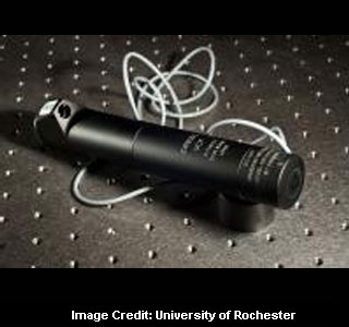
Offering a clear, high-resolution, 3D image of what lays below the surface within a span of few seconds, the prototype device is more affordable and time-friendly than the traditional method. The newly developed technology apparently uses a liquid lens setup which is made up of a droplet of water that takes the place of the glass in a standard lens. Once the electrical field around the water droplet changes, the droplet presumably modifies its shape and hence, alters the focus of the lens.
The liquid lens seemingly empowers the device to take thousands of pictures focused at different depths below the skin’s surface. Combining these images may produce a fully in-focus image of all of the tissue up to 1 millimeter deep in human skin. The newly introduced technology also employs near infrared light instead of ultrasounds. Replacing ultrasounds with near infrared light can possibly aid in generating images that are precise with micron-scale resolution instead of a millimeter-scale resolution.
“My hope is that, in the future, this technology could remove significant inconvenience and expense from the process of skin lesion diagnosis,” added Professor Jannick Rolland. “When a patient walks into a clinic with a suspicious mole, for instance, they wouldn’t have to have it necessarily surgically cut out of their skin or be forced to have a costly and time-consuming MRI done. Instead, a relatively small, portable device could take an image that will assist in the classification of the lesion right in the doctor’s office.”
Apparently, the developed device was tested in in-vivo human skin with positive results. Experts will be conducting further investigations to examine the efficacy of this device in a clinical research environment for distinguishing between different types of lesions. The research findings seem to have a great significance in the medical section.
The research was presented at the 2011 annual meeting of the American Association for the Advancement of Science in Washington, D.C., on February 19.