
 If you’re watching the complex processes in a living cell, it is simple to miss something important, particularly if you are observing changes that take a long time to unfold and require high-spatial-resolution imaging. However, a latest research seems to make it achievable to examine activities that take place over hours or even days inside cells. This novel research may potentially solve various mysteries associated with molecular-scale events occurring in these tiny living things.
Researchers are believed to have found a method of using nanoparticles to illuminate the cellular interior in order to reveal these slow processes. They were from the National Institute of Standards and Technology (NIST) and the National Institute of Allergy and Infectious Diseases (NIAID).
Nanoparticles are known to be thousands of times smaller than a cell and have a range of applications. One type of nanoparticle called a quantum dot apparently glows when exposed to light. These semiconductor particles could possibly be coated with organic materials which are modified to be attracted to definite proteins within the part of a cell.
NIST lead researcher and biophysicist, Jeeseong Hwang said that, “Quantum dots last longer than many organic dyes and fluorescent proteins that we previously used to illuminate the interiors of cells. They also have the advantage of monitoring changes in cellular processes while most high-resolution techniques like electron microscopy only provide images of cellular processes frozen at one moment. Using quantum dots, we can now elucidate cellular processes involving the dynamic motions of proteins.â€
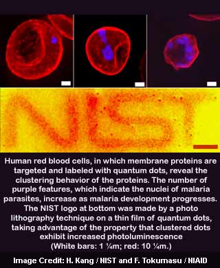
For their latest research, the team appears to have focused mainly on characterizing quantum dot properties, differentiating them with other imaging techniques. In one instance, they were noted to have utilized quantum dots designed to target a specific type of human red blood cell protein. Moreover, these quantum dots supposedly, form part of a network structure in the cell’s inner membrane.
When these proteins come together in a healthy cell, the network seems to provide mechanical flexibility to the cell. This mechanical flexibility could perhaps further squeeze via narrow capillaries and other tight spaces. However, when the cell gets infected with the malaria parasite, the structure of the network protein may change.
NIAID biophysist Fuyuki Tokumasu said that, “Because the clustering mechanism is not well understood, we decided to examine it with the dots. We thought if we could develop a technique to visualize the clustering, we could learn something about the progress of a malaria infection, which has several distinct developmental stages.â€
The findings revealed that as the membrane proteins collect, the quantum dots attached to them appear to be encouraged in order to gather themselves and glow more brightly. This may possibly permit the researchers to watch as the clustering of proteins progresses.
Hwang further said, “Some concerns remain over toxicity and other properties. But altogether, our findings indicate that quantum dots could be a valuable tool to investigate dynamic cellular processes.â€
More largely, the team found that when quantum dots attach themselves to other nanomaterials, the dots’ optical properties apparently change in distinctive ways in every case.
The findings also revealed that quantum dot optical properties could perhaps be altered as the nanoscale environment changes. This may offer better prospect of using quantum dots to sense the local biochemical environment inside cells.