
Just some time back, we reported about a distinct laser probe that seemingly showed the potential to detect oral cancer early. Initial detection and timely treatment may aid in boosting survival rate among individuals with this form of cancer. Apparently experts from the University of Sheffield and Sheffield Teaching Hospitals NHS Foundation Trust are developing a novel test for identifying oral cancer in the early stages itself. A dentist can possibly conduct this test by gathering cells from a patient’s mouth with the help of a brush.
It is anticipated that the test can give out a precise diagnosis in less than 20 minutes for lesions suspected of oral cancer. Present day test for detecting oral cancer seemingly includes extraction of scalpel. Then a biopsy and off-site laboratory tests will be conducted on the scalpel. However, this test appears very time-consuming. The latest test simply requires the removal of cells with a brush, placing them on a chip, and inserting the chip into the analyser. It will simply take 8-10 minutes. Time waste on cutting and making a number of trips to the dentists can be saved. Even the cost for detecting oral cancer seems to be minimized.
Professor Martin Thornhill Professor of Oral Medicine at the University of Sheffield and a Consultant in Oral Medicine at Sheffield Teaching Hospitals and the lead investigator, remarked, “This new affordable technology will significantly increase our ability to detect oral cancer in the future. Diagnosis currently involves removing a small piece of tissue from the mouth and sending it to a pathologist. This is typically done at a hospital, can take a week or more and involve extra visits for the patient. With the new technology, a brush would be used to painlessly remove a few cells from the lining of the mouth that would be analysed within minutes in the presence of the patient, so that the patient would know the result before leaving the clinic.”
Investigators have already triggered clinical trials on patients at Charles Clifford Dental Hospital for two years. They are testing if the technology provides accurate results and are aiming to make it as sensitive as possible. Once approval is achieved on the usage of this technology, experts will be carrying out a biopsy so that it can become a daily application at dentist surgeries in the future. It was ascertained that with early identification of oral cancer, patients can be treated sooner and can improve a five-year survival rate of more than 90 percent.
Professor Thornhill enlightened, “This technology will make it easier for us to screen suspicious lesions in the mouth and separate non-cancerous lesions from those where there is a risk of cancer and those where cancer has already developed. We have just started to recruit patients to a study that is designed to ensure that the new technology is at least as good as the old method at distinguishing these different types of lesion. Ultimately, dentists and doctors may be able to use this technology to check suspicious lesions in the mouth and reassure the vast majority of patients that they haven´t got cancer without even having to send them to the hospital.”
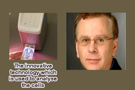
Since oral cancer is difficult to recognize in the early stages itself, the overall survival rate is only around 50 percent which is among the lowest rates for all major cancers. The technology employs the latest techniques in microchip design, nanotechnology, microfluids, image analysis, pattern recognition and biotechnology. It does this to possibly curb several crucial functions of a state-of-the-art clinical pathology laboratory onto a nano-bio-chip the size of a credit card. Nano-bio-chips are disposable and selected like a credit card into a battery-powered analyser.
Once a brush-biopsy sample is achieved it can be placed on the card and microfluidic circuits will wash cells from the sample into the reaction chamber. All the cells pass through mini-fluidic channels that are about the size of small veins and come in contact with ‘biomarkers.’ It was mentioned that the biomarkers react only with certain types of diseased cells. Regions of the cells and cell compartments are possibly lit up due to the LEDs, or light-emitting diodes employed by the machine. The way they glow in response to the LEDs possibly indicates whether the cells are healthy or diseased. The technology may also be beneficial in diagnosing and managing heart attacks, diabetes and other diseases.