
Technology has certainly gained momentum. While we’ve seen innumerable iPhone applications that help in combating various health problems from depression to patient monitoring, smartphones it appears could also play a key role in radiology. A new study has apparently discovered that smartphones may probably permit fast diagnosis of acute appendicitis from a remote location.
With the help of a handheld device or mobile phone, radiologists, the study reveals may now be able to appropriately diagnose acute appendicitis from a far away places. This could be possible courtesy distinct software incorporated in the device.
A medical emergency needing the surgical removal of the organ, appendicitis or even inflammation and infection of the appendix could be really painful. If left undiagnosed or untreated there are high chances of the inflamed appendix rupturing. This could probably be fatal as it may cause toxins to spill into the abdominal cavity. Though appendicitis could occur at any age, as per the National Institutes of Health, it is found to be most prevalent between ages 10 and 30.
“The goal is to improve the speed and accuracy of medical diagnoses, as well as to improve communications among different consulting physicians,” remarked the study’s lead author, Asim F. Choudhri, M.D., fellow physician in the Division of Neuroradiology at Johns Hopkins University in Baltimore. “When we can make these determinations earlier, the appropriate surgical teams and equipment can be assembled before the surgeon even has the chance to examine the patient.”
Experts suggest that a typical patient arriving at the emergency room with suspected appendicitis will apparently undergo computed tomography (CT) along with a physical examination. Diagnosis may be delayed if the radiologist or special consultant is not available at that moment. This could be mainly due to the immediate unavailability for CT images interpretation, consequently augmenting the chances of rupture. On the upside if the images could be transmitted over a mobile device, they could allow for instant consultation and diagnosis even from a remote location. Radiologists say that this could help in proper planning of surgery.
“This new technology can expedite diagnosis and, therefore, treatment,” Dr. Choudhri mentioned.
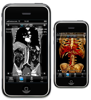
At the University of Virginia in Charlottesville, as part of the study, CT examinations of the abdomen and pelvis of 25 patients with pain in the right lower abdomen were reviewed. These were analyzed over an encrypted wireless network by five radiologists armed with an iPhone G3 that was bundled with the OsiriX Mobile medical image viewing software. To confirm whether or not they had appendicitis, all the patients had gone through surgical confirmation or follow-up evaluations.
“The scans can be read in full resolution with very little panning, and the software allows the reader to zoom and adjust the contrast and brightness of the image,” Dr. Choudhri said. “The radiologist is evaluating actual raw image data, not snapshots.”
The experts observed that 15 of the 25 patients were accurately recognized as having acute appendicitis on 74 (99 percent) of 75 interpretations, with one false negative. With no false positive readings, in eight of the 15 patients who had appendicitis, calcified deposits within the appendix were appropriately identified in 88 percent of the interpretations. In 96 percent of interpretations, all 15 patients showed apparent signs of inflammation close to the appendix that were correctly identified. In addition to this 10 of the 15 had fluid near the appendix, which was again correctly identified in seemingly 94 percent of the interpretations. All five readers supposedly could also identify three abscesses correctly.
“The iPhone interpretations of the CT scans were as accurate as the interpretations viewed on dedicated picture archiving and communication system (PACS) workstations,” Dr. Choudhri added.
With a hope to enhance patient outcomes, Dr. Choudhri further elucidates that before any handheld mobile device could be considered practical for clinical use, patient privacy concerns would need to be addressed. He however suggests that this method appears to have great potential for improving emergency room care.
The study was presented at the annual meeting of the Radiological Society of North America (RSNA).