
 UT Southwestern Medical Centre claims to be the first site in North Texas to launch the next generation in computed tomography CT scanners. This device allows doctors to view an entire organ in less than a second. It also tracks blood flow through the brain or to a tumor. All this is done with less radiation exposure to patients. Aquilion One dynamic volume computed tomography (CT) is capable of forming a detailed 3-D movie of an organ in real time. Aquilion One which was manufactured by Toshiba exposes patients to less radiation.
CT specifically helps in early detection of strokes and heart attacks. Diagnostic speed plays a critical factor in survival and recovery of a patient. For patients, the new technology can mean less time in a scanner and less exposure to radiation, said Dr. Phil Evans. Its great speed may also mean less required contrast materials. It can also benefit patients who have difficulty remaining still, such as children, the elderly and trauma patients.
“Dose has been a concern in the medical literature for a long time and people have been very concerned about it,†said Dr. Phil Evans, associate vice president for clinical imaging and professor of radiology who directs UT Southwestern’s Clinical Imaging Services. “One of the great things about this is that you can do a scan with about half the radiation dose and half the contrast media, so the dose is less and the image is better.â€
The machine’s technology is capable of taking continuous or intermittent images. Hence UT Southwestern radiologists expect better visualization in neurology, trauma, whole body, lung, cardiac, vascular and pediatric studies. Various other functions provided by the machine include distinctive capabilities in orthopedic and joint studies, diagnosing renal function, and even vocal-cord analysis.
“This has the potential to impact the daily medicine we currently practice and help us identify future clinical pathways,†Dr. Evans said. “UT Southwestern is fortunate to have clinical experts and forward-looking researchers who will really be the ones to determine its best uses. Technology, even the best technology, still depends on the expertise of those using it.â€
It is observed that other scanners join together strips of images to assimilate a complete picture by using 4, 16, 32 or 64 slice machines. This is because one strip covers a larger area which requires less time and swaths overall. According to published analyses it is observed that radiation can be reduced by as much as 80 percent.
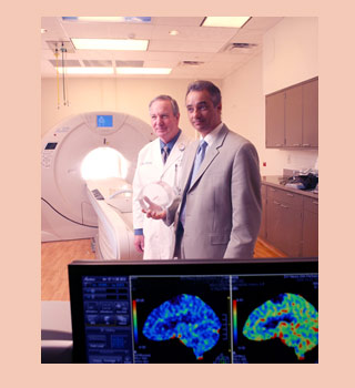
“One of the most exciting things about this technology is the real-time ability to image changing anatomy,†said Dr. Phillip Purdy, professor of radiology and neurological surgery. “We have been able to image physiology such as blood flow in parts of the brain, but now we can image the entire brain faster and more safely.â€
This new device uses 320 high resolution X-ray detectors in each rotation. It takes about one third of a second for the 320 slice machine. Where as other scanners take 12 to 15 seconds to complete. UT Southwestern physicians anticipate to accurately diagnosing a stroke or heart attack in about 20 minutes. It will also gauge tissue damage.
In order to confirm a heart attack doctors perform tests like EKG, CT angiography, nuclear testing and catheterization which take hours or even days to complete. Other clinical practices include patients who can’t get an MRI due to the presence of a pacemaker, or vocal-cord analysis capturing a patient phonating. The machine provides more flexibility and helps to accurately position patients with trauma or disabilities and is sturdy enough to accommodate obese patients.
UT Southwestern physicians expect that the Aquilion One device may be valuable in many of the medical center’s unique research projects. The capability of the machine to move backward and forward in time through the images helps the researchers to analyze properly the effects of tissue damage or vascular flow.