
 Cardiologists and surgeons subjecting patients to pacemakers, bypass surgery or angioplasties can possibly benefit from the following discovery. Experts from The University of Western Ontario have supposedly introduced a novel imaging technique that provides a single, 3-D high-resolution image of the heart showing its vasculature and presence of scar tissue within the muscle. This new tool designed to improve a patient’s outcome was performed with a 3-Tesla MRI.
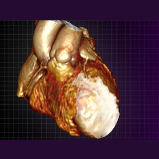
It is known that injuries to the heart like heart attacks or viral inflammation generally result in permanent damage or scarring of its muscle. This technique may first give a 3-D coronary image that is acquired by continuous infusion of contrast known as gadolinium. It is believed that gadolinium makes the blood-pool light up brightly. Once the image is obtained, 3-T MRI takes it and provides a high resolution, 3-D image of the heart showing coronary blood vessels.
Experts affirm that scar tissue is slow to give up this contrast agent. So its signal may be retained in spite of washing out contrast from the blood stream and normal tissues. After 20 minutes, a repeat image may be performed that highlights the heart’s scar, also in 3-D. Both the images are supposedly taken in the identical way with the help of a similar MRI pulse sequence. Therefore the images are accurately suited to be fused on one another. The ultimate result appears to be a fused, 3-D model of the heart showing the heart’s vessels as well as scar tissue.
Dr. James White remarked, “We’ve known for some time that myocardial (heart) scar tissue can be imaged using MRI, but what we’ve now been able to do is to take this imaging to another level. This is the first time we have been able to visualize myocardial scar and the heart’s blood vessels at the same time. We are able to construct a three dimensional model of a person’s heart to immediately understand the relationship between the heart’s blood vessels and related permanent injury. This will help direct surgeons and cardiologists to better target the blood vessels that lead to muscle capable of responding to their therapy, rather than to muscle that is irreversibly diseased.â€
The novel imaging technique was performed by the researchers on 55 patients. These patients were referred for either bypass surgery or a specialized pacemaker designed to improve heart function called Cardiac Resynchronization Therapy (CRT). The experts were supposedly able to prove that this technique helps in planning vascular-based cardiac interventions. Before conducting bypass or angioplasty procedures, surgeons may have to decide whether or not to open up blocked blood vessels. Since they can view a scar in that region, no benefit will possibly be expected. In a similar manner CRT pacemaker delivered to regions of scarred heart muscle can avoid any assistance from this therapy.
The research was published on-line in the Journal of the American College of Cardiology: Cardiovascular Imaging.