
 Philips Medical Systems has unveiled a revolutionary piece of medical equipment that shows human organs, bones and even blood vessels in living patients. Called the Brilliance iCT machine, this new type of an X-ray scanner provides in-depth three-dimensional images of the inner workings of the human body.
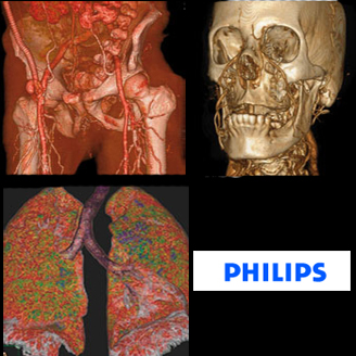
Using the Brilliance iCT machine, the patient’s organs like the heart and lungs appear as of they exist outside the patient. Besides, remarkable images of the human skeleton have also been captured using this new medical scanner.
Besides providing a 3D view of the human anatomy, the Brilliance iCT Scanner also offers reduces scanning time and thus, this reduces the dose of radiation that the patient receives by 80% as well.
Philips newly launched Brilliance iCT scanner works really fast and is able to capture an image of a human organ such as the heart in the time it takes the organ to beat just twice.
Basically, the scanner takes large numbers of X-rays and then feeds them into a computer, which then produces an image that can be rotated and viewed from different directions. Cross-sections through that part of the body being scanned also help to produce the final image.
According to Steve Rusckowski, CEO of Philips Medical Systems, “We are seeking to make a difference in how radiologists can prevent, diagnose, treat and monitor disease and allow them to focus more on their patients.”
“This scanner allows radiologists to produce high-quality images and is also designed to reduce patients’ exposure to X-rays. It is so powerful it can capture an image of the entire heart in just two beats while also including technology that has reduced radiation doses by up to 80%,” he added.
The Philips Brilliance iCT Scanner was unveiled at the annual meeting of the Radiological Society of North America in Chicago on November 25, 2007.