
 Understanding tumor cells seems to be the most accurate way of developing cancer treatments. But the absence of tumor models appears as a major hindrance in cancer research. Heeding to this, a team of experts has now crafted a 3-D model that displays more accurate tests of new cancer therapies.
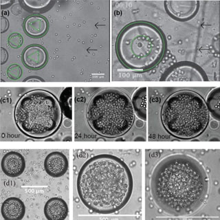
Some three years ago, the team had come up with a low-cost, easy fabrication technique to make spherical cavities in PDMS (polydimethylsiloxane), a widely used silicon organic polymer. And now researchers have found that these cavities can be utilized as a scaffolding to grow numerous tumor spheroids. This in turn may serve as realistic models for cancer cells.
“Basically, any laboratory that works with cells could adopt our new spherical microcavity system to do their own 3-D experiments or drug screening on hundreds or even thousands of little tumor spheroids,” said team leader Michael R. King, Ph.D., of Cornell University.
According to this research, the three-dimensional spheroids can fasten cancer drug discovery by providing a realistic and easily accessible substrate on which to test drugs. The 3-D nature appears as an asset because in the body, tumor cells grow in 3-D–yet most laboratory studies of cancer have been done in 2-D. The new 3-D tumor spheroids can reportedly provide a realistic tumor oxygen environment that cues the blood vessel growth which nourishes tumors.
The research is published in the current issue of Biomicrofluidics, a publication of the American Institute of Physics.