
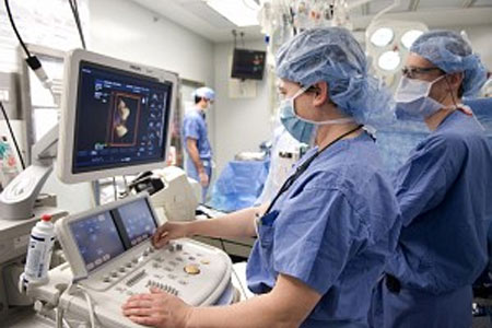
The health world has now stumbled upon a novel technology that keeps a track on critically ill patients. Vanderbilt cardiothoracic anesthesiologists and surgeons have introduced a tool called Transesophageal echocardiography (TEE) that efficiently supervises those undergoing cardiac surgery and are critically ill. TEE appears as a diagnostic procedure that includes feeding an ultrasound probe through a patient’s mouth and into their esophagus to analyze heart function.
Since the esophagus is close to the heart, this tool can seemingly produce high-resolution images as the organ pumps. It may be extremely vital for locating cardiac blood clots, masses and tumors and can detect the severity of valve problems, congenital heart diseases as well as aortic tears. The newly crafted ultrasound tool was employed to monitor cardiac patients in ICUs both before and after surgery. As a result, TEE seemingly allowed medical staff to quickly intervene in a crisis and use the resulting information to make crucial treatment decisions.
“The ability to monitor cardiac function and filling in a serial fashion with miniaturized, disposable TEE probes leads to optimal patient outcome and appropriate resource utilization. The only real barrier to the routine use of this technology is physician training,†said Chad Wagner, M.D., a cardiothoracic anesthesiologist and director of the Cardiovascular Intensive Care Unit (CVICU).
The study subjects were trained to use a TEE simulator that combines a photo-realistic, three-dimensional computer-generated model and generate a simulated ultrasound image to help users visualize and understand complex cardiac anatomy. A disposable TEE simulator was used to care for a 66-year-old patient who became unstable in the ICU following open heart surgery. It was suggested that a miniaturized disposable TEE probe I can diagnose and treat the cause of the patient’s low blood pressure.
The novel TEE supposedly has major implications in the medical section.