
 This news apparently promises to provide enhanced identification and monitoring of Alzheimer’s disease. Researchers at the University of California, San Diego have supposedly created a quick and precise technique for quantifying delicate, sub-regional brain volume loss via magnetic resonance imaging (MRI).
By using the methods to the recently finished dataset of the multi-institution Alzheimer’s disease Neuroimaging Initiative (ADNI), the researchers supposedly illustrated that such sub-regional brain volume measurements could surpass accessible measures for trailing seriousness of Alzheimer’s disease, counting extensively utilized cognitive testing and measures of worldwide brain-volume loss.
The universal design of brain atrophy ensuing from Alzheimer’s disease has apparently been known for a long time through autopsy studies. But utilizing this information toward precise diagnosis and monitoring of the disease has supposedly only lately been made feasible by enhancements in computational algorithms that could mechanize recognition of brain structures with MRI.
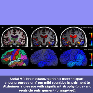
The new techniques supposedly offer swift recognition of brain sub-regions pooled with measures of alteration in these areas across time. The methods apparently need at least two brain scans to be conducted on the same MRI scanner over duration of numerous months. The new research supposedly illustrates those alterations in the brain’s memory areas, a region of the temporal lobe known as the entorhinal cortex, apparently provides receptive measures of the initial stages of the disease.
Anders M. Dale, PhD, professor of neurosciences and radiology at the UC San Diego School of Medicine, who led the research, commented, “Loss of volume in the hippocampus is a consistent finding when using MRI, and is a reliable predictor of cognitive decline. However, we have now developed and validated imaging biomarkers to not only track brain atrophy, but distinguish the early stages of Alzheimer’s disease from changes related to normal aging.â€
Research co-author James Brewer, MD, PhD, a neurologist and assistant professor in the Departments of Radiology and Neurosciences at UC San Diego, mentioned, “The technique is extremely powerful, because it allows a researcher to examine exactly how much brain-volume loss has occurred in each region of the brain, including cortical regions, where we know the bad proteins of Alzheimer’s disease build up. We are particularly excited to use the techniques in new clinical trials, but also to reexamine old clinical trial data where global measures of brain shrinkage were applied. These new findings suggest that such global measures are less sensitive than regional measures for detecting the changes specific to Alzheimer’s disease – the changes these drugs are targeting.â€
The scientists at various locations across the U.S. apparently examined almost 300 patients with slight cognitive impairment, around 169 healthy controls and about 129 subjects suffering from AD and then gauged rates of sub-regional cerebral volume alteration for every group. Power calculations were claimed to be conducted to recognize areas that could offer the most sensitive result measures in clinical trials of disease-modifying agents.
The research was published in the Proceedings of the National Academy of Science (PNAS).