
 Technology is advancing by leaps and bounds. While last week we saw the development of synthetic red blood cells that mimic the functions of natural ones, here’s a new lab chip that appears to steer the behavior of heart cells.
Biomedical engineers from the Johns Hopkins in collaboration with colleagues from Seoul National University Korea claim to have developed a laboratory chip that boasts of nanoscopic grooves and ridges. This is supposedly capable of growing cardiac tissue that behaves very similar to natural heart muscle.
The researchers reveal that the heart cells cultured in this manner astonishingly utilized a nanosense that garnered instructions. This included chalk talks for growth and function acquired from the physical patterns on the nanotextured chip alone. Apparently no special chemical cues were used to guide the tissue development in distinct ways. Researchers are of the opinion that this tool may find place in the fabrication of novel therapies or diagnostic tests for cardiac disease.
Reflecting an important enhancement for scientists to culture cells in the lab, this new technique will allow them to gain m ore information about cardiac disorders along with potential remedies.
“Heart muscle cells grown on the smooth surface of a Petri dish, would possess some, but never all, of the same physiological characteristics of an actual heart in a living organism,†mentioned Andre Levchenko, a Johns Hopkins associate professor of biomedical engineering at the Whiting School of Engineering.
“That’s because heart muscle cells—cardiomyocytes—take cues from the highly structured extracellular matrix or ECM, which is a scaffold made of fibers that supports all tissue growth in mammals. These cues from the ECM influence tissue structure and function, but when you grow cells on a smooth surface in the lab, the physical signals can be missing. To address this, we developed a chip whose surface and softness mimic the ECM. The result was lab-grown heart tissue that more closely resembles the real thing,†he further added.
Levchenko further shared when he and his colleagues analysed the natural heart tissue taken from a living animal, they ‘immediately noticed that the cell layer closest to the extracellular matrix grew in a highly elongated and linear fashion. The cells orient with the direction of the fibers in the matrix, which suggests that ECM fibers give structural or functional instructions to the myocardium, a general term for the heart muscle.’
According to Levchenko, these instructions seem to have been delivered on the nanoscale. As part of the novel development Levchenko and his Korean colleagues, working with Deok-Ho Kim, a biomedical engineering doctoral student from Levchenko’s lab and the lead author of the PNAS article, created a two-dimensional hydrogel surface. This surface appeared to have simulated the rigidity, size and shape of the fibers found all through a natural ECM network. Besides, it is claimed to have been bio-friendly and tailored from nontoxic polyethylene glycol that displays an array of long ridges.
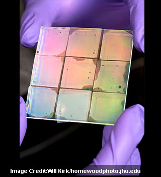
With the ridges resembling the folded pattern of corrugated cardboard, the ridged hydrogel was housed upon a glass slide about the size of a U.S. dollar coin. In this manner a number of different types of chips with ridge widths spanning from 150 to 800 nanometers, groove widths ranging from 50 to 800 nanometers, and ridge heights varying from 200 to 500 nanometers were developed. This allowed the experts to gain control of the surface texture over more than five orders of magnitude of length.
“We were pleased to find that within just two days, the cells became longer and grew along the ridges on the surface of the slide,†further commented Kim.
The researchers observed an enhanced coupling between neighboring cells just like the architecture of the natural layers of heart muscle tissue. With cells that were grown on smooth, unpatterned hydrogels were found to have remained smaller and a little less organized. They also seemed to show poorer cell-to-cell coupling between layers.
“It was very exciting to observe engineered heart cells behave on a tiny chip in two dimensions like they would in the native heart in three dimensions,†Kim remarked.
“Perhaps most surprisingly, these tissue functions and the structure of the engineered heart tissue could be controlled by simply altering the nanoscale properties of the scaffold. That shows us that heart cells have an acute ‘nanosense,’†Levchenko mentioned further. “This nanoscale sensitivity was due to the ability of cells to deform in sticking to the crevices in the nanotextured surface and probably not because of the presence of any molecular cue,â€
Levchenko believes that engineering surfaces that seem to exhibit identical nanoscale features in three dimensions instead of just two could offer a much better way for control of the structure and function of cultured heart tissue.
The experiments using this technique have been described in this week’s online Early Edition of Proceedings of the National Academy of Sciences.